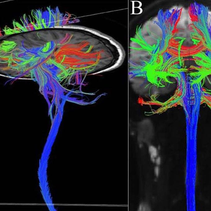


Tractography in the brain and spinal cord
DTI and Tractography have found numerous clinical applications in spinal cord (trauma, MS, inflammation, tumors) with the ability to detect certain pathologic processes at an early stage, guide optimal therapy and predict outcomes. Standard SS-EPI DWI is difficult to used in spinal cord due to area's inhomogeneities. Newer techniques (such as Siemens' RESOLVE and ZOOMit EPI and GE's FOCUS) can achieve reduced TE, reduced ESP and smaller FOV, which can lead to higher image quality. Images show an example of tractography in the brain and spinal cord. Image A shows a sagittal view of tractography, with an axial slice of anatomical data centered in the brain. The white box at the top and bottom of the image represent the seed points, that is, where the tractography starts and end (deterministic algorithm). Image B shows the same subject with coronal orientation. Images were acquired with a 64-channel coil and a readout-segmented (RESOLVE) sequence (Keil et al., 2013).

