


MRI Technologistalmost 9 years ago
MR Imaging of brain's meningioma
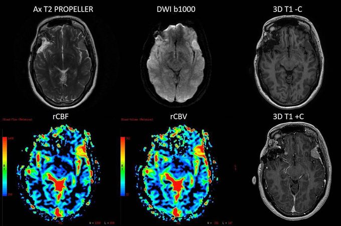
Multiparametric MR Imaging of brain's meningioma, based on high-resolution morphological (axial T2 PROPELLER and IR-prepared 3D T1 GRE pre and post contrast are shown here) and functional (DWI b1000 and DSC PWI are shown here, also DCE PWI was acquired) acquisitions. The high rCBV and rCBF of the lesion should be noted.
Other commentsSign in to post comments. You don't have an account? Sign up now!
