


MRI Technologistabout 9 years ago
MR Imaging of the prostate gland
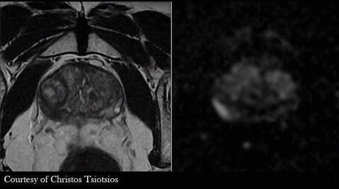
High-resolution, multiparametric (anatomical, functional and quantitative) MR Imaging of the prostate gland. Figure shows a lesion on the peripheral zone with low signal on axial T2-w FSE, high signal on DWI b1400 and low signal on corresponding ADC map (not shown here), findings that indicate malignancy.
Other commentsSign in to post comments. You don't have an account? Sign up now!
