


MRI Technologistalmost 9 years ago
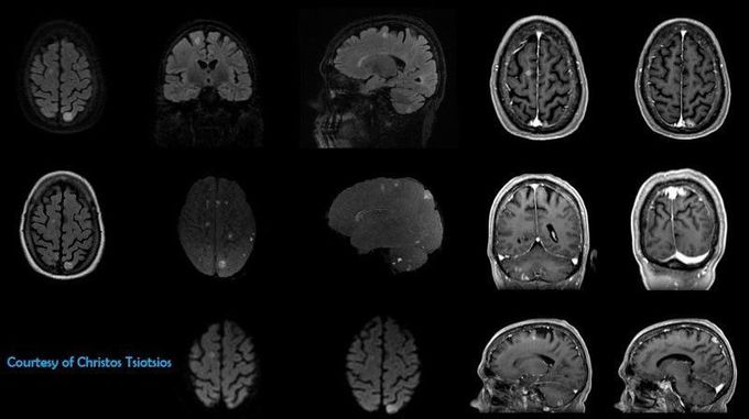
Multiple brain metastases
Figure shows a patient with multiple brain metastases. High resolution MR imaging based on 3D T2 FLAIR with fat-suppression, 3D T1 FSPGR, 2D axial FLAIR and axial DWI (b0, b500, b1000). Moreover, 3D MIPs of the volumetric T2 FLAIR, for segmentation and volumetric calculation of the metastatic lesions, are shown.
Other commentsSign in to post comments. You don't have an account? Sign up now!
