


MRI Technologistalmost 9 years ago
3D ultrasound and magnetic resonance imaging
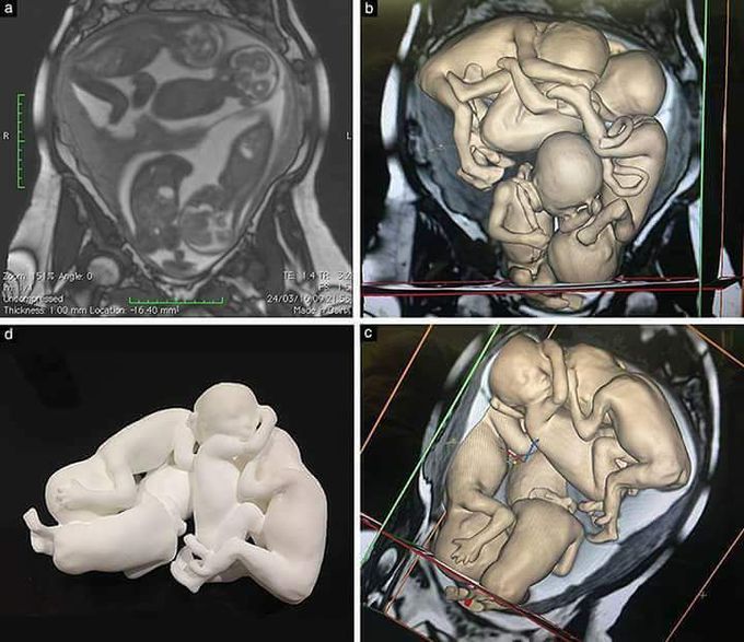
In a new UOGJournal article, Werner et al. describe how they used data from three-dimensional (3D) ultrasound and magnetic resonance imaging (MRI) in a monochorionic diamniotic quadruplet pregnancy to produce 3D virtual and physical models. (a) MRI (T2-weighted true fast imaging with steady-state precession sequence) in monochorionic diamniotic quadruplet pregnancy at 26 weeks. Data from MRI were used to construct 3D mathematical virtual model (b,c) and 3D physical model (d). http://ow.ly/JIlg30dgQfA Become an ISUOG Journal member for access: http://www.isuog.org/Membership/
Other commentsSign in to post comments. You don't have an account? Sign up now!
