


MRI Technologistalmost 9 years ago
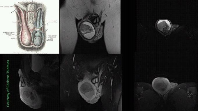
High-resolution scrotum MR Imaging
Traditionally, due to its low cost, ready availability, and proved diagnostic accuracy, US has been the primary imaging modality for the evaluation of scrotal disease. However, US is limited by its relatively small useful field of view, operator dependence and inability to provide much information on tissue characterization. Magnetic resonance (MR) imaging, with its excellent soft-tissue contrast and good spatial resolution, is increasingly being used as both a problem-solving tool in patients who have already undergone US and as a primary modality for the evaluation of suspected disease.
Other commentsSign in to post comments. You don't have an account? Sign up now!
