


MRI Technologistalmost 9 years ago
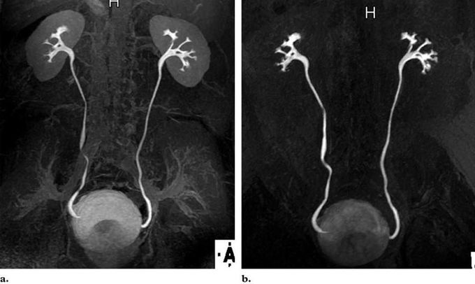
Comparison of different sequences and flip angles used for excretory MR urography
(a) On a coronal MIP image from excretory MR urographic data obtained with a 3D interpolated fat-suppressed gradient-echo sequence (VIBE/THRIVE/LAVA) during breath holding, soft-tissue suppression is minimized owing to the use of a relatively low flip angle of 12°. (b) Coronal MIP image from excretory MR urographic data obtained with a 3D gradient-echo MR angiographic sequence shows improved background tissue suppression due in part to the use of a higher flip angle of 40° (Leyendecker et al 2008)
Other commentsSign in to post comments. You don't have an account? Sign up now!
