


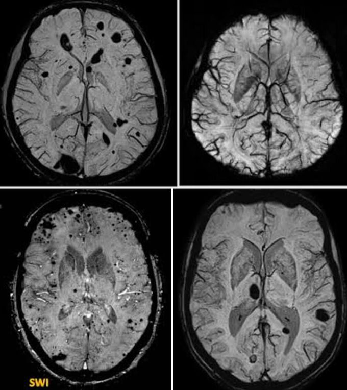
Susceptibility-weighted imaging (SWI)
Susceptibility-weighted imaging (SWI) is a neuroimaging technique, which uses tissues magnetic susceptibility differences to generate a unique contrast, different from that of proton density (PD), T1, T2, and T2*. SWI uses a fully flow/velocity compensated, RF spoiled, high-resolution, three-dimensional (3D) gradient recalled echo (GRE) scan. Both the magnitude and phase images are saved. The phase image is high pass (HP) filtered to remove unwanted artifacts (background field inhomogeneities caused by air-tissue interfaces and main magnetic field effects). The magnitude image is then combined with the HP filtered phase image to create an enhanced contrast magnitude image referred to as the susceptibility weighted image (SWI). It is also common to create minimum intensity projections (minIP) over 8 to 10 mm to better visualize vein connectivity.
