


MRI Technologistalmost 9 years ago
Magnetic Resonance Imaging (MRI)
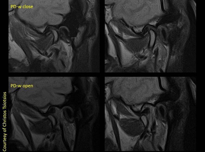
Magnetic Resonance Imaging (MRI) is the most useful and widely used imaging modality for the evaluation of the temporomandibular joint (TMJ). Compared with computed tomography (CT) and arthrography, MRI provides better tissue contrast for the visualization of soft tissue and other articular structures of the TMJ. Direct visualization of the articular disc by MRI without the use of any contrast medium is a distinct advantage over arthrography. Appropriate selection of coils and proper imaging techniques play a major role in TMJ imaging. Images courtesy of Christos Tsiotsios.
Other commentsSign in to post comments. You don't have an account? Sign up now!
