


Diffusion-Weighted MR Imaging of the head and neck
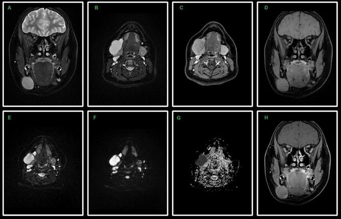
Diffusion-Weighted MR Imaging of the head and neck using Read-out Segmented Echo-Planar Imaging technique. 14-year-old female patient with submandibular tumor. Coronal T2-w FSE Dixon water image (A), axial T2-w FSE Dixon water image (B), axial post-contrast T1-w FSE Dixon water image (C), coronal T1-w FSE Dixon water image (D), axial RS-EPI DWI with b0 (E), b1000 (F) and ADC map (G) and coronal post-contrast T1-w FSE Dixon water image (H). RS-EPI technique provides homogeneous, high-quality images with reduced susceptibility artifacts and geometric distortions in the head and neck area. Submandibular tumor shows high signal intensity on the high-b-value image (F) and low signal intensity on the corresponding ADC map (G), which indicates malignancy. Image dataset was acquired at 3.0 Tesla, Siemens Skyra. Courtesy of Bac Nguyen.
