


MRI Technologistover 8 years ago
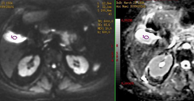
Fig (left) Diffusion-weighted image (b = 600 s/mm2) and (right) ADC map. Figure shows T2 "shine through" effect. In the DWI, the gallbladder is hyperintense, not because it restricts diffusion but because it has a long T2 relaxation time. It is verified with the ADC map where it is also hyperintense. The take-home point is that Trace DW images are both diffusion- and T2-weighted. Lesions that have very long T2-values may appear bright even though they do not restrict diffusion. #MRI #diffusion #DWI
Other commentsSign in to post comments. You don't have an account? Sign up now!
