


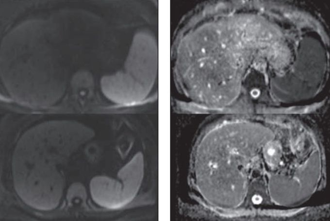
Fig 52-year-old man with chronic hepatitis C
Fig 52-year-old man with chronic hepatitis C without evidence of fibrosis at liver biopsy (stage F0; top) and 67-year-old woman with cirrhosis secondary to chronic hepatitis C (stage F4; bottom). Single-Shot Echo-Planar Diffusion-Weighted Image (SS-EPI DWI) with b = 700 (left) mm2/s and apparent diffusion coefficient (ADC) map (right) (using b = 0 and 700 mm2/s) are shown. In patient without fibrosis, hepatic ADC was within normal range, measuring 1.6 × 10⁻³ s/mm2, with liver appearing brighter than spleen (which is known to have low ADC). In patient with fibrosis, hepatic ADC was decreased, reaching spleen ADC, measuring 1.0 × 10⁻³ s/mm2 (Taouli et al. 2009). Bi-exponential models (eg IVIM) may provide better results due to the high vascularity of liver tissue.

I was diagnosed as a Hepatitis B carrier in 2015, with early signs of liver fibrosis. At first, antiviral medications helped control the virus but over time, resistance developed, and the effectiveness faded. I began to lose hope. In 2021, I discovered NaturePath Herbal Clinic despite my skepticism, I decided to give their herbal treatment a try.To my surprise, after just six months, my blood tests came back negative for the virus.It was nothing short of life-changing.I never expected such incredible results from a natural treatment. But it not only cleared the virus it restored my hope, my health, and my peace of mind.If you or someone you know is battling Hepatitis B, I truly encourage you to explore the natural healing path offered by NaturePath Herbal Clinic. It gave me a second chance and it might do the same for you.www.naturepathherbalclinic.com info@naturepathherbalclinic.com
