


Cerebral cavernous venous malformations MRI
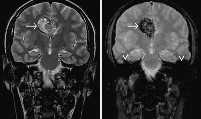
Cerebral cavernous venous malformations, commonly known as cavernous haemangioma or cavernoma, are common cerebral vascular malformations, usually with characteristic appearances on MRI. MRI is the modality of choice, demonstrating a characteristic “popcorn” or "berry" appearance with a rim of signal loss due to hemosiderin, which demonstrates prominent blooming on susceptibility weighted sequences. T1 and T2 signal is varied internally depending on the age of the blood produces and small fluid-fluid levels may be evident. Gradient echo or T2* sequences are able to delineate these lesions better than T1 or T2 weighted images. In patients with familial or multiple cavernous angiomas GRE T2* sequences are very important in identifying the number of lesions missed by conventional Spin echo sequences. Note the susceptibility artifact (arrowhead) on T2*-w (right image) due to local inhomogeneities.
