


MRI Technologistalmost 9 years ago
Study finds gadolinium hot spots from MRI contrast, but no clinical effects
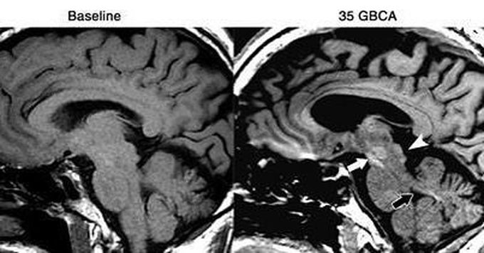
Fig. T1-weighted MRI of a 61-year-old man with left frontal glioblastoma shows high signal intensity in the colliculi (arrowhead), red nucleus and substantia nigra (white arrow), and superior cerebellar peduncle (black arrow) after 35 GBCA administrations, compared with baseline MRI prior to GBCA. Images courtesy of Radiology.
Other commentsSign in to post comments. You don't have an account? Sign up now!
