


MRI Technologistabout 9 years ago
DWI
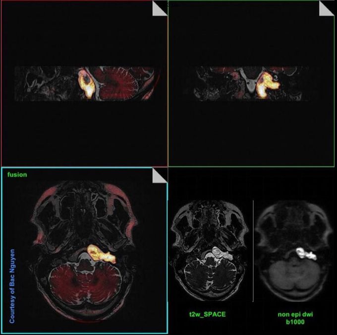
DWI is a useful technique for the evaluation of cholesteatomas. It can be used to detect them when the physical examination is difficult and CT findings are equivocal. Moreover, it is especially useful in the evaluation of recurrent cholesteatoma. Fig. Image fusion --> High resolution T2-w SPACE (modified 3D TSE) sequence, which provides us anatomical information, with a HASTE (single-shot TSE) DWI b1000, which provides us functional information. Non-EPI DWI assists in detection and evaluation of the lesion. Image dataset acquired at Siemens Aera 1.5 Tesla, courtesy of Bac Nguyen.
Other commentsSign in to post comments. You don't have an account? Sign up now!
