


Rectal cancer MR Protocol and planning
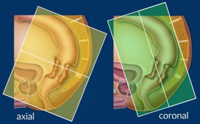
High resolution 2D T2-weighted fast spin echo sequences in the sagittal, axial and coronal plane are required for state-of-the-art staging of rectal cancer. The slice thickness should be 3 mm. Gadolinium-enhanced MR does not improve diagnostic accuracy and is not included in the protocol. Start with the sagittal series. These can be used to plan the axial images (left), perpendicular to the rectal wall at the level of the tumor to avoid volume averaging (yellow box). Coronal images (right) are planned parallel to the anal canal (green box), especially in low-rectal tumors in order to accurately evaluate the depth of tumor invasion into the anal sphincter. The cranial border of the field of view (FOV) is vertebral body L5, the caudal border is below the anal canal. Finally, Diffusion weighted imaging (DWI) can be useful for tumor and lymph node detection in primary staging.
