


MRI Technologistalmost 9 years ago
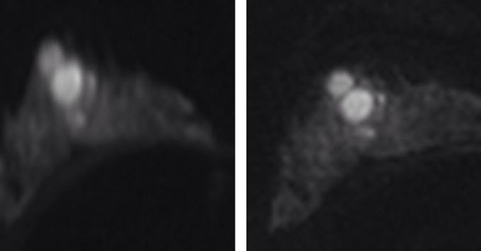
DW images with b = 850 sec/mm2 obtained with two different DW imaging methods, (left) single-shot echo-planar image, and (right) readout-segmented echo-planar image in 24-year-old woman with breast cancer (fibroadenoma). Significantly stronger geometric distortion artifacts can be seen on single- shot echo-planar image compared with readout-segmented echo-planar image. Readout-segmented echo-planar images provide significantly higher anatomic detail than single-shot echo-planar images because of reduced T2* blurring and reduced TE, ESP and encoding times.
Other commentsSign in to post comments. You don't have an account? Sign up now!
