
MRI Technologistalmost 9 years ago

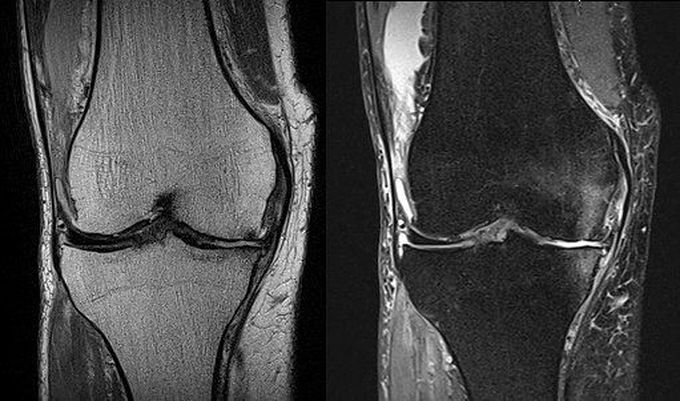
Fat suppression is an important technique in musculoskeletal MR imaging to improve the visualization of bone marrow lesions. Figure: Coronal PD-W TSE (left) and fat suppressed PD-W TSE (right) MR images of the right knee. Fat suppressed PD-W image can visualize the bone marrow edema on the medial condyle of femur and tibia, while no lesion is highlighted on the PD-W image. Image dataset acquired at 1.5 Tesla. Reference: Images courtesy of Bac Nguyen
Other commentsSign in to post comments. You don't have an account? Sign up now!

