


MRI Technologistover 8 years ago
Susceptibility-weighted imaging
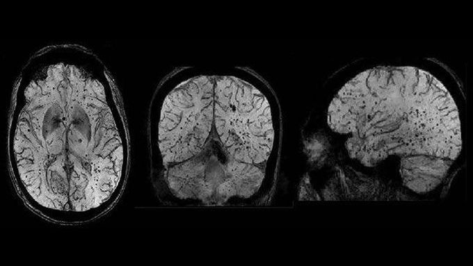
Susceptibility-weighted imaging (SWI) is a neuroimaging technique, which uses tissues magnetic susceptibility differences to generate a unique contrast, different from that of proton density (PD), T1, T2, and T2*. SWI can be used better at higher field strengths. First of all, magnetic susceptibility increases accordingly to the square of the magnetic field strength. Moreover, the high signal-to-noise (SNR) ratio available at higher magnetic fields allows higher resolution scans. Finally, stronger magnetic fields allow shorter echo times (TE) without a loss of contrast which can reduce scan time and motion related artifacts.
Other commentsSign in to post comments. You don't have an account? Sign up now!
