


MRI Technologistalmost 9 years ago
The time-of-flight or in-flow effect
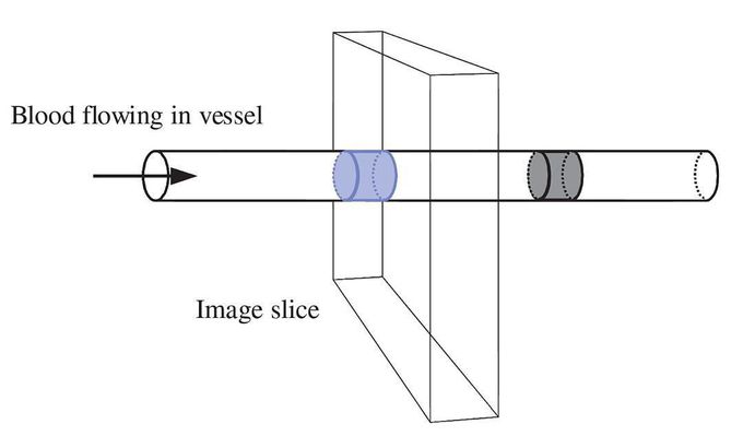
The blood vessel is shown crossing through the imaging slice. When the sequence is repeated, the previously excited blood (coloured grey) has moved on and the bolus within the slice (coloured blue) has fully relaxed magnetization M0.
Other commentsSign in to post comments. You don't have an account? Sign up now!
