


MRI Technologistalmost 9 years ago
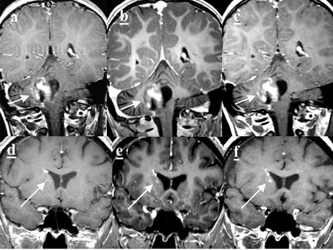
a, d: 2D FSE T1-weighted; b, e: BRAVO (MP-RAGE); c, f: T1 CUBE (SPACE/ VISTA). TOP ROW: The 2D FSE T1W and T1 CUBE images give superior depiction of the margins of this enhancing cerebellar tumor, in part because of greater CNR of contrast-enhancing tumor to adjacent cerebellar white matter. BOTTOM ROW: A channel of this venous angioma is best appreciated with the T1 CUBE, because of greater CNR relative to BRAVO and probably because of smaller section thickness relative to 2D FSE T1W.
Other commentsSign in to post comments. You don't have an account? Sign up now!
