


MRI Technologistabout 9 years ago
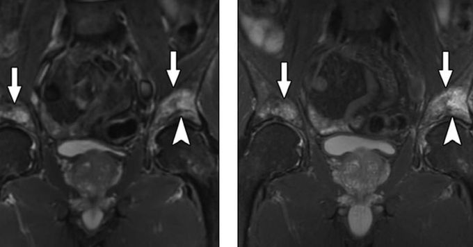
(left) Coronal STIR and (right) coronal T2W FSE Dixon (water image) of pelvis reveal high-signal edema within both acetabuli (arrows). There is associated linear area of low signal within right acetabulum (arrowhead), which suggests presence of insufficiency fracture. Improved SNR ratio and higher resolution of Dixon image allow better visualization of trabecular detail. Both techniques provide robust fat suppression. (Gerdes et al. 2007)
Other commentsSign in to post comments. You don't have an account? Sign up now!
