


Intravoxel incoherent motion (IVIM)
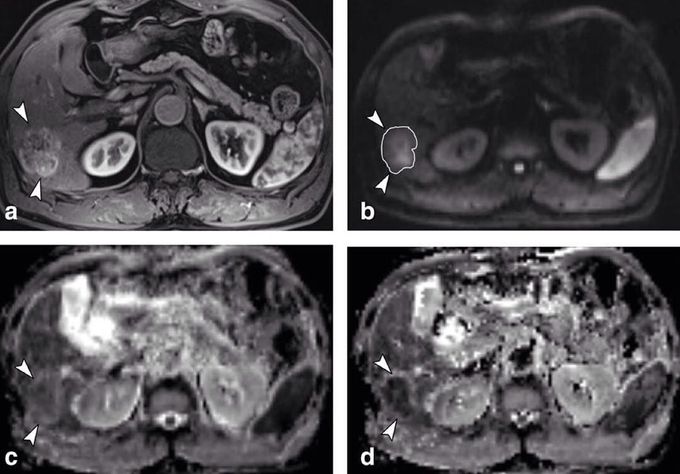
Intravoxel incoherent motion (IVIM) imaging is a concept and a method initially introduced and developed by Le Bihan et al. to quantitatively assess all the microscopic translational motions that could contribute to the signal acquired with diffusion MRI. In biological tissues, these motions essentially are molecular diffusion of water and microcirculation of blood in the capillary network (perfusion). The concept introduced by D. Le Bihan is that water flowing in randomly oriented capillaries (at the voxel level) mimics a random walk ("pseudo-diffusion"). According to IVIM theory, the fast component of diffusion (represented by D*) is related to microperfusion (mainly on low b values, 0-100 s/mm2), whereas the slow component (represented by D) is linked to pure molecular diffusion. Owing to this bi-compartmental model, the signal decay of IVIM DWI was described using a bi-exponential model. Using IVIM-based analysis, it is possible to derive quantitative indexes that describe conventional ADC (ADCtotal), tissue water diffusivity (pure diffusion/ D), tissue perfusion (pseudodiffusion coefficient/ D*), and the perfusion fraction of tissues (f), which can also be displayed as parametric maps. Because there are four fitted parameters, a minimum of four b values are needed to characterize bi-exponential signal attenuation, though this choice is not recommended. A larger number of b values would provide more data support for the estimates and would enable parameter uncertainties to be evaluated, which is not possible with four b values. IVIM model can be used on clinical applications in the liver, pancreas, prostate and other well-perfused body organs. Figure shows a 3.2 cm surgically confirmed HCC in 59-year-old man. The mass (arrowheads) showed heterogeneous enhancement during arterial phase (a) and high signal intensity on DWI (b factor, 800 sec/mm2) (b). The tumor showed a slightly lower ADC value (arrowheads) than surrounding liver parenchyma on ADC map (c). True D was obviously lower (arrowheads) than liver parenchyma on D map (d) (Yoon et al. 2013.
