


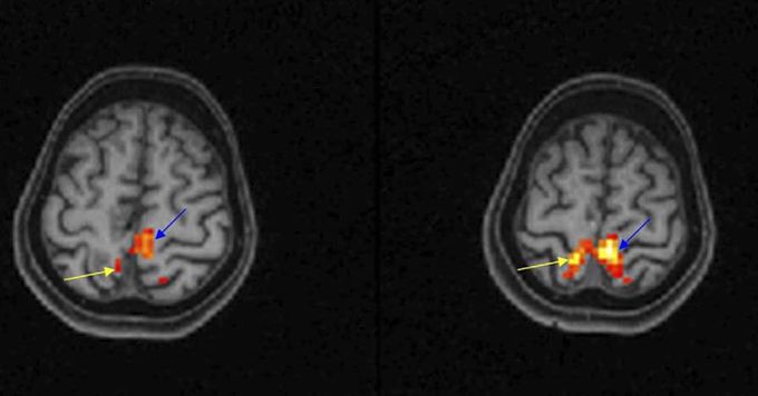
Functional magnetic resonance imaging (fMRI) provides a sensitive, non-invasive tool for mapping patterns of activation in the working human brain. The most common #fMRI technique is the blood oxygenation level dependent (#BOLD) technique. This technique measures localized changes in tissue oxygen content by alterations in the local magnetic field owing to the different magnetic properties of oxygenated (diamagnetic) and deoxygenated (paramagnetic) blood. The basic technique for a clinical fMRI scan involves acquisition of high-resolution 3 dimensional anatomic images (eg MPRAGE or T2), followed by BOLD GRE or SE EPI images showing functional activation. Current fMRI research concentrates on task-related studies and designs measuring functional connectivity. Task-related activation studies aim to characterize the neural responses to experimental conditions, which may be visualized as maps of brain activity. Figure shows axial T1-W images in neurologic format with overlaid functional activation show right foot (yellow arrow) and left foot (blue arrow) activation in the parasagittal precentral gyri. The patient was asked to curl his toes during "on task" and rest during "off task" (L.M. Shah et al, Seminars in Roentgenology 2010).
