


Pathologic conditions of the distal biceps brachii tendon
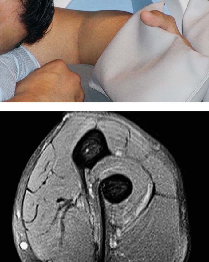
Pathologic conditions of the distal biceps brachii tendon are of clinical interest, with partial and complete tears being the most common. However, the anatomy of the distal biceps brachii tendon makes imaging of the distal tendon somewhat difficult. An innovation in patient positioning for MRI of the distal biceps tendon was described in which the patient lies prone with the arm overhead, the elbow flexed to 90°, and the forearm supinated, so that the thumb points superiorly. The acronym FABS (Flexed elbow, ABducted shoulder, forearm Supinated) has been used to describe this position. The FABS position creates tension in the tendon and minimizes its obliquity and rotation, resulting in a “true” longitudinal view of the tendon. Imaging with FABS positioning can complement conventional_MRI, especially in the axial plane, in the assessment of the distal biceps tendon.
