


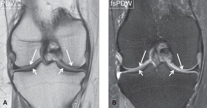
#MR_imaging is the method of choice to identify articular cartilage injuries and evaluate the post-operative reconstitution of cartilage repair tissue. Although various biochemical techniques (such as T2 mapping, T1rho mapping, dGEMRIC, CEST, sodium MRI) are becoming feasible for the assessment of cartilage tissue architecture, conventional morphologic #MRI remains the mainstay for the pre- and post-operative evaluation of the articular cartilage in clinical practice. Most experience and good results in morphological imaging of cartilage and subchondral pathology were gathered with (i) 2D PDW FSE with intermediate TE value (30–60 ms) and high ETL (8-10), (ii) 3D DESS (ideal FA for maximum cartilage signal is 20°-30° and for highest CNR between cartilage and synovial fluid is 90°) and (iii) 3D spoiled gradient-echo (SPGR). Fat suppression in these sequences was found useful to better visualize cartilage pathology, increase CNR and reduce chemical shift artifacts. Coronal images (A, B) of the knee demonstrate a normal articular cartilage (arrows). #MRI #MRI_of_cartilage
