


MRI Technologistalmost 9 years ago
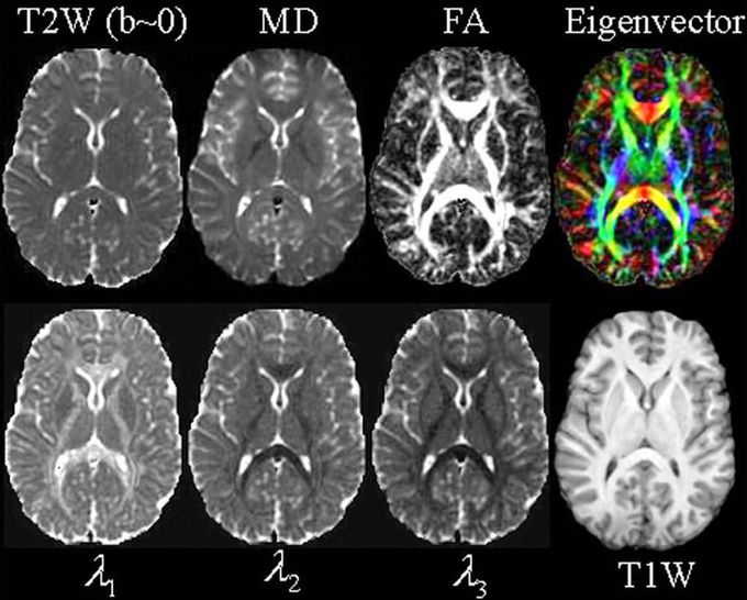
Quantitative maps from a #DTI experiment. The images include the T2-weighted (T2W) “reference” (or b=0) image from DTI data; the mean diffusivity (MD - note similar contrast to T2W image with CSF appearing hyperintense); fractional anisotropy (FA - hyperintense in white matter); the major eigenvector direction indicated by color (red = R/L, green = A/P, blue = S/I) weighted by the FA (note that specific tract groups can be readily identified); the major, medium and minor eigenvectors (11, 12, and 13, respectively). A conventional T1-weighted (from a 3D MP-RAGE) at the same anatomical location is also displayed. #MRI
Other commentsSign in to post comments. You don't have an account? Sign up now!
