


Medshotsabout 2 years ago
Ruptured venule
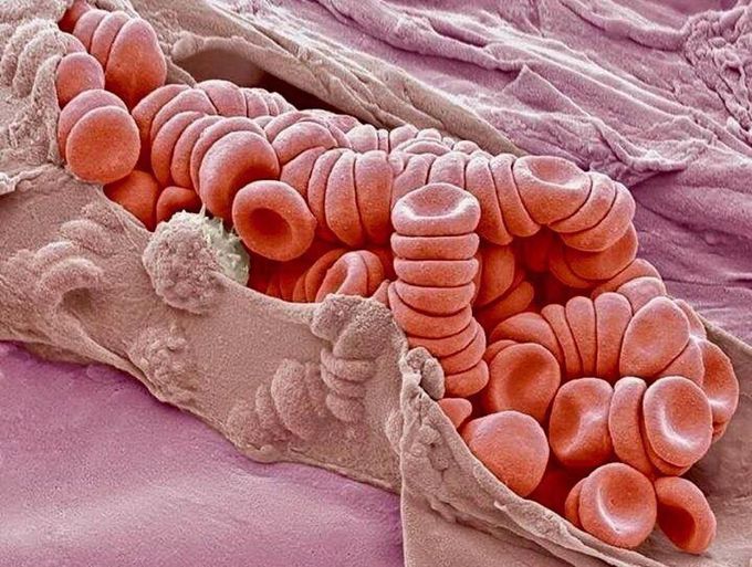
Wow, this is how a ruptured venule looks like under a magnified microscope.Shown here is a colored scanning electron micrograph (SEM) showing stacks (rouleaux) of red blood cells exposed inside a torn venule.A venule is a very small blood vessel in the microcirculation that allows deoxygenated blood to return from the capillary beds to the larger blood vessels (veins). Red blood cells are the most abundant cell in the blood. They have no nucleus and are about 7 micrometers across. Magnification: x2300 when printed at 10 centimetres wide.
Other commentsSign in to post comments. You don't have an account? Sign up now!
Related posts
Practice questions regarding microscopeTypes of light microscopesScanning Electron Microscope(SEM)Microscope practicalCause of RuptureLeft Ventricular RuptureDiagnosis of Achilles’ Tendon Rupture during a Virtual ExaminationRuptured bladder linked to shinglesHistoplasmosisRuptured eardrum
If you get a hole in your tympanic membrane, it’s called a ruptured eardrum. (Your eardrum separates your outer ear from your middle ear.) Infection, trauma, loud sounds or foreign objects in your ears can cause a ruptured eardrum. In most cases, a ruptured eardrum will heal on its own in a few weeks. But sometimes, it requires surgical repair, such as tympanoplasty.
