


Dr.Sana4 months ago
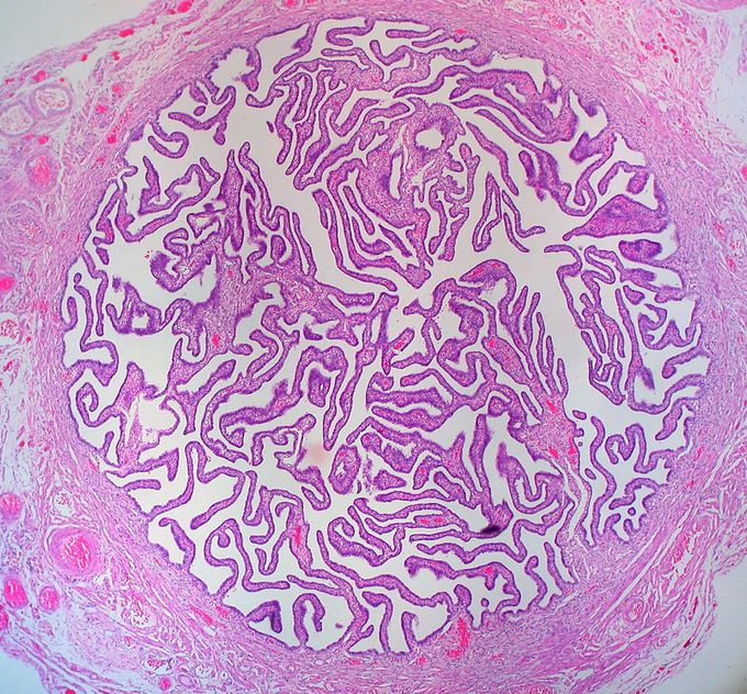
Histology of FALLOPIAN TUBE
The cross section of Fallopian tube shows four distinct layers: serosa, subserosa, lamina propria and innermost mucosal layer. The serosa is derived from visceral peritoneum. Subserosa is composed of loose adventitious tissue, blood vessels, lymphatics, an outer longitudinal and inner circular smooth musclecoats. This layer is responsible for the peristaltic action of the Fallopian tubes. Lamina propria is a vascular connective tissue. There are two types of cells within the simple columnar epithelium of the Fallopian tube (oviduct). Ciliated cells predominate throughout the tube, but are most numerous in the infundibulum and ampulla. Estrogen increases the production of cilia on these cells.
Other commentsSign in to post comments. You don't have an account? Sign up now!
Related posts
What does amniocentesis test for?When is amniocentesis performed?Road traffic accidentHello dear colleague
I have a question.
Is curtage of uterus(for placenta residue) out of abdoman is standard procedure? I mean uterus contact with skin epidermis of abdoman.
Thanks for your attention. I’m looking forward to your reply.Why is the limit for spontaneous abortion 20 weeks, what happens at 20th week? Why we define it as spontaneous abortion up to 20 week and why not after?Heart failureHave a great Wirk𝙼𝚢𝚘𝚖𝚎𝚌𝚝𝚘𝚖𝚒𝚎𝚜 𝚙𝚎𝚛 𝚌é𝚜𝚊𝚛𝚒𝚎𝚗𝚗𝚎If your uterus is mean to you and you know it, clap your hands.
👏 👏
If your uterus is mean to you and you know it, clap your hands.
👏 👏
If your uterus is mean to you, and you really wanna show it, if your uterus is mean to you, clap your hands.
👏 👏Preparation for USMLE
