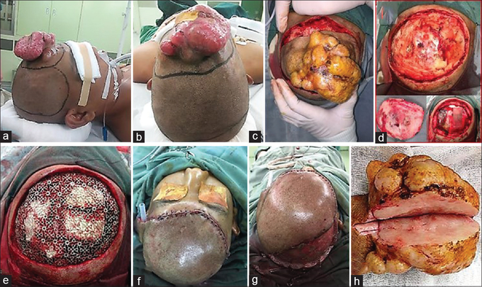
Cranioplasty and reconstruction
(a-c) A wide radical excision with 4 cm distance from neoplasm margin and translational skin flap line was drawn. (d) After wide excision was performed, calvarial defect was observed without involvement of duramater below; as frontal bone craniectomy was performed. (e) A bone defect 10 × 8cm was made and cranioplasty using titanium mesh was placed. (f) The skin defect was then reconstructed by translational skin flap and (g) split-thickness skin graft. (h) Mass with an irregular, skin colored, multiple lobulated mass, 12 × 10 × 5 cm in diameter was excised As in histopathological findings, using hematoxylin and eosin (HE) stained sections showed a densely cellular and poorly circumscribed tumor in the dermis layer, comprising interwoven bundles and fascicles of uniform spindle shaped cells arranged in a “storiform” or “cartwheel” pattern The tumor cells had monotonous appearance with oval nuclei, vesicular chromatin, inconspicuous nucleoli, and scanty-to-moderate cytoplasm. A panel of immunohistochemistry (IHC) comprising CD-34 and vimentin was applied.